1. Method for separating spectrally overlapping multiphoton upconverted emission bands through spectral power dependence analysis
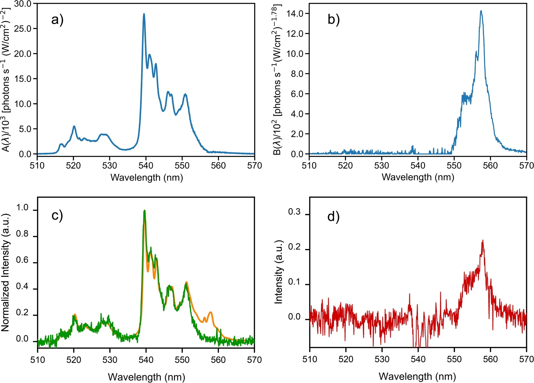
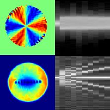
The high quantum efficiency and long lifetimes of upconverting rare-earth-doped systems make luminescence band spectral overlap a particularly common phenomenon, frequently associated with various excitation and relaxation pathways. While it is often considered challenging to experimentally distinguish such overlapping luminescent bands, herein it is proposed a new method based on the different spectral responses of those luminescent lines to the excitation power at a single pumping wavelength laser. A model for the population of the overlapping bands in terms of the pump laser power is considered and fitted for each measured wavelength, resulting in separated spectra for each pathway. The results obtained with the proposed method are considered with direct excitation measurements, showing the effectiveness and simplicity of the method in comparison with more traditional approaches.
Journal of Luminescence 257, 119685 (2023).
2. Observation and analysis of nonlinear scattering using the scattered-light imaging method
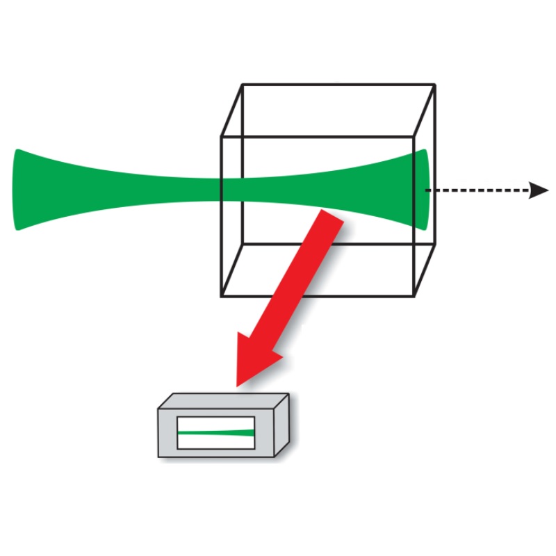
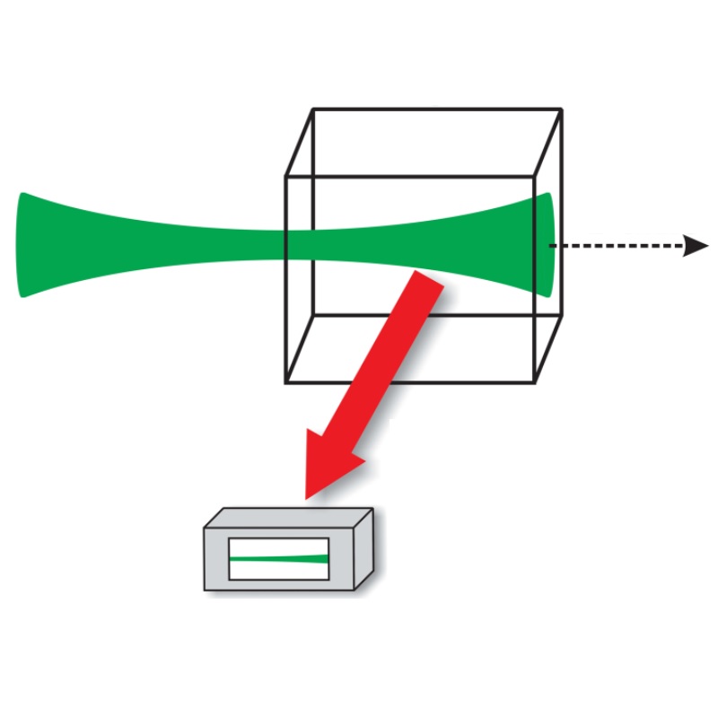
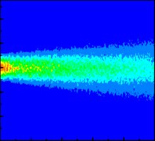
Optical phenomena like nonlinear absorption and nonlinear refraction are often determined through light transmittance techniques. However, in turbid or resonant media, scattering may also significantly attenuate the laser beam, such that absorptive losses may be overestimated. Thus, while transmittance techniques are suitable to determine the power extinction, it is often difficult to characterize the contribution of nonlinear scattering in a sample. In this work we applied the scattered-light imaging method to discriminate the nonlinear extinction due to nonlinear absorption and nonlinear scattering contributions in a turbid medium constituted of TiO2 nanoparticles suspended in acetone. The single-shot method collects an image of the beam transverse profile evolution along the propagation axis inside the sample. It was observed that while the results obtained by the transmittance technique indicate the extinction coefficient (including scattering and absorption contributions), the results obtained with the scattered-light imaging method discriminate the contribution of each phenomenon. Therefore, the results of both approaches are complementary and allow a more complete characterization of turbid media.
Physical Review A 105, (2022).
3. 2D Thermal Maps Using Hyperspectral Scanning of Single Upconverting Microcrystals: Experimental Artifacts and Image Processing
Whereas lanthanide-based upconverting particles are promising candidates for several micro- and nanothermometry applications, understanding spatially varying effects related to their internal dynamics and interactions with the environment near the surface remains challenging. To separate the bulk from the surface response, this work proposes and performs hyperspectral sample-scanning experiments to obtain spatially resolved thermometric measurements on single microparticles of NaYF4: Yb3+,Er3+. Our results showed that the particle's thermometric response depends on the excitation laser incidence position, which may directly affect the temperature readout. Furthermore, it was noticed that even minor temperature changes (<1 K) caused by room temperature variations at the spectrometer CCD sensor used to record the luminescence signal may significantly modify the measurements. This work also provides some suggestions for building 2D thermal maps that shall be helpful for understanding surface-related effects in micro- and nanothermometers using hyperspectral techniques. Therefore, the results presented herein may impact applications of lanthanide-based nanothermometers, as in the understanding of energy-transfer processes inside systems such as nanoelectronic devices or living cells.
ACS Applied Materials &$\mathsemicolon$ Interfaces 14, 38311--38319 (2022).
4. Second harmonic scattering of redox exfoliated two-dimensional transition metal dichalcogenides
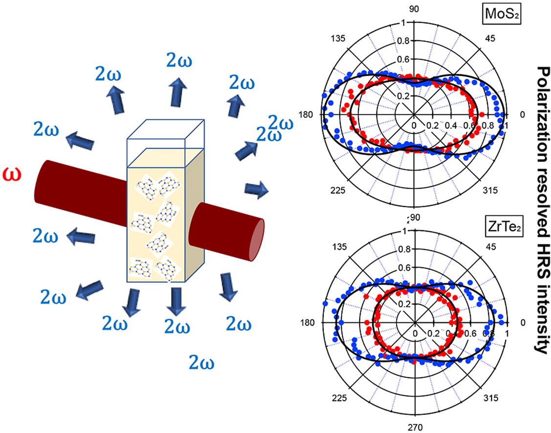
We report the second harmonic scattering experiments from acetonitrile liquid suspensions of two-dimensional metal dichalcogenide nanoflakes,: the semiconducting MoS2 and WS2, the metallic NbS2, and the semi-metallic ZrTe2. Because the study is directly performed in the liquid phase, no symmetry breaking can be attributed to a supporting substrate as it is often the case in the investigation of the second harmonic generation efficiency for these materials. A comparative look of the origin of the nonlinear response using polarization analysis of the hyper Rayleigh scattering intensity is performed. Retardation is clearly exhibited by all nanoflakes considering their average dimensions but remains weak. This is attributed to the non-negligible role played by the nanoflake edges or the presence of surface absorbed polyoxometalates (POMs), in all cases.
Optical Materials 133, 112780 (2022).
5. Influence of the surrounding medium on the luminescence-based thermometric properties of single Yb3+/Er3+ codoped yttria nanocrystals
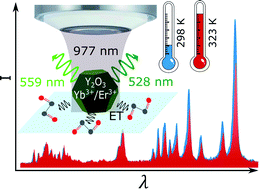
While temperature measurements with nanometric spatial resolution can provide valuable information in several fields, most of the current literature using rare-earth based nanothermometers report ensemble-averaged data. Neglecting individual characteristics of each nanocrystal (NC) may lead to important inaccuracies in the temperature measurements. In this work, individual Yb3+/Er3+ codoped yttria NCs are characterized as nanothermometers when embedded in different environments (air, water and ethylene glycol) using the same 5 NCs in all measurements, applying the luminescence intensity ratio technique. The obtained results show that the nanothermometric behavior of each NC in water is equivalent to that in air, up to an overall brightness reduction related to a decrease in collected light. Also, it was observed that the thermometric parameters from each NC can be much more precisely determined than those from the "ensemble" equivalent to the set of 5 single NCs. The "ensemble" parameters have increased uncertainties mainly due to NC size-related variations, which we associate to differences in the surface/volume ratio. Besides the reduced parameter uncertainty, it was also noticed that the single-NC thermometric parameters are directly correlated to the NC brightness, with a dependence that is consistent with the expected variation in the surface/volume ratio. The relevance of surface effects also became evident when the NCs were embedded in ethylene glycol, for which a molecular vibrational mode can resonantly interact with the Er3+ ions electronic excited states used in the present experiments. The methods discussed herein are suitable for contactless on-site calibration of the NCs thermometric response. Therefore, this work can also be useful in the development of measurement and calibration protocols for several lanthanide-based nanothermometric systems.
Nanoscale Advances 3, 6231--6241 (2021).
6. Microcontroller-based magnetometer using a single nitrogen-vacancy defect in a nanodiamond
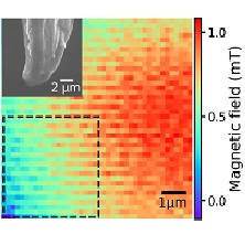
The measurement of magnetic properties of various physical systems with nanometric spatial resolution raises in-terest in areas as materials science, biotechnology and information storage and processing. In the present work amicrocontroller-based magnetometer was built using a single nitrogen-vacancy defect in a nanodiamond. The imple-mented nanomagnetometry method is simple and relies on the frequency modulation of the nitrogen-vacancy defectelectron spin resonance using square pulses of an externally applied magnetic field and employs a single microwavesource. The developed system has a reasonable sensitivity of 4$μ$T/√Hz and is able to measure magnetic field varia-tions in time around 4 mT/s. This system was used for nanoimaging the inhomogeneous spatial magnetic field profileof a magnetized steel microwire, and a spatial magnetic field gradient of 13$μ$T/ 63 nm was measured. Besides itsusefulness for nanoscale imaging of magnetic fields, the present work can be of interest in the development of compactnanodiamond based magnetometers.
AIP Advances 10, 025323 (2020).
7. Observation and analysis of creation, decay, and regeneration of annular soliton clusters in a lossy cubic-quintic optical medium
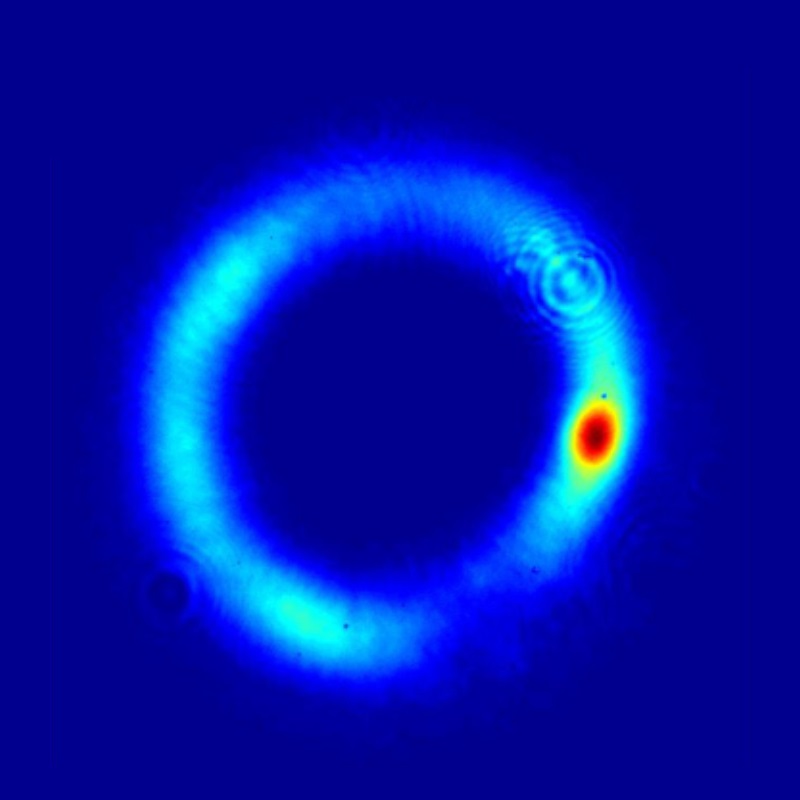
We observe and analyze formation, decay, and subsequent regeneration of ring-shaped clusters of (2+1)-dimensional spatial solitons (filaments) in a medium with cubic-quintic (focusing-defocusing) self-interaction and strong dissipative nonlinearity. The cluster of filaments, which remains stable over $≈$ 17.5 Rayleigh lengths, is produced by the azimuthal modulational instability from a parent ring-shaped beam with embedded vorticity $l=1$. In the course of still longer propagation, the stability of the soliton cluster is lost under the action of nonlinear losses. The annular cluster is then spontaneously regenerated due to power transfer from the reservoir provided by the unsplit part of the parent vortex ring. Thus, a secondary interval of the robust propagation of the regenerated cluster is identified. The experiments use a laser beam (at wavelength 800 nm), built of pulses with temporal duration 150 fs, at the repetition rate of 1 kHz, propagating in a cell filled by liquid carbon disulfide. Numerical calculations, based on a modified nonlinear Schrödinger equation which includes the cubic-quintic refractive terms and nonlinear losses, provide results in close agreement with the experimental findings.
Phys. Rev. A 102, 033523 (2020).
8. Toward single-shot characterization of nonlinear optical refraction, absorption, and scattering of turbid media
Disordered media are often susceptible to optical damage even at low irradiation doses, since the correspond-ing microscopic structure may suffer from processes such as bleaching, chemical reactions, or morphologicalchanges. The disorder often introduces linear and nonlinear (NL) light scattering, and currently it is a challengeto perform single-shot characterization of NL refraction, NL absorption, and NL scattering using the morecommon NL transmittance techniques. This work discusses how the scattered light imaging method and D4σ techniques can be combined to characterize the NL optical constants of thick turbid media. Therefore, it shouldhelp researchers aiming to draw a detailed physical picture of turbid samples at the single laser shot level.
Phys. Rev. A 102, 033503 (2020).
9. Influence of strong light beams on the nonlinear refraction and absorption coefficients of transparent materials
We explore the effective nonlinear (NL) optical response of transparent materials illuminated by strong light beams. Two different setups were used to study the influence of the numerical aperture (NA) corresponding to the collecting lens on the measured coefficients in the open- and closed-aperture Z-scan transmittance. For that, we have reproduced experiments in CS_2 at 532 nm in the picosecond regime using the D4σ-Z-scan method. It is found that measurements of high-order NL coefficients are not possible after a limit defined by the self-focusing of light inside the cell, and that stimulated light scattering is not significant to explain alone the saturation behavior that occurs in the NL refraction measurements. Indications are given for future Z-scan-based experiments that may clarify the boundary conditions to be respected for the measurement.
J. Opt. Soc. Am. B 36, 3411--3416 (2019).
10. Effective model for nonlinear refraction and extinction coefficients in the presence of stimulated light scattering
We present in this paper a wave-coupled model (WCM) to describe the effective nonlinear (NL) optical response of transparent materials illuminated by strong light beams in conditions such that stimulated light scattering is relevant. At low intensities, the WCM indicates that the NL scattering behaves similarly to a third- and fifth-order polarization susceptibility. However, at sufficiently high intensities, NL light scattering will lead to highly nonperturbative effective NL refraction and NL absorption coefficients. This is an important remark, given that attempts to explain NL scattering contributions as a perturbative NL response from data obtained at low intensities will become discrepant from the data obtained at high intensities. As an application of the model, we show that the existing divergent interpretations of the carbon disulfide (CS2) NL response in the picosecond and femtosecond regimes by several authors are consistent with the WCM, which indicates a direction for future experiments that may clarify the present controversy.
J. Opt. Soc. Am. B 35, 2977--2985 (2018).
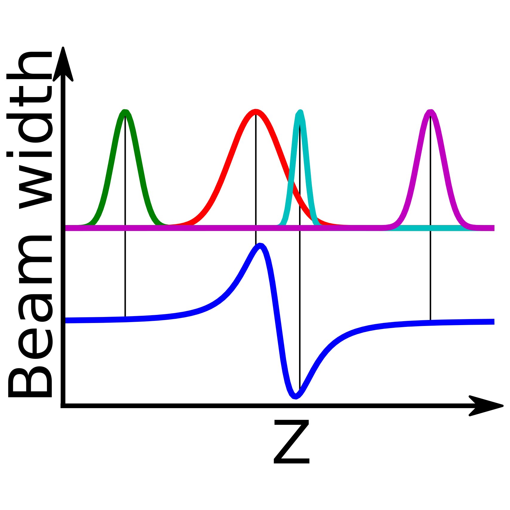
11. D4$σ$ curves described analytically through propagation analysis of transverse irradiance moments
The far-field changes in the beam width of an intense light beam propagating inside optical materials are investigated aiming the measurement of the nonlinear properties of the samples. The beam width is characterized in this Letter in terms of the transverse irradiance moments (TIMs) that allow an accurate description of the beam interaction and propagation. TIMs are rigorously related to the paraxial beam propagation parameters, and it is also possible to consider elliptical and astigmatic beams. Experimental data with very good signal-to-noise ratio were obtained for the reference materials carbon disulfide and fused quartz.
Opt. Lett. 41, 2081--2084 (2016).
12. Measurements of the nonlinear refractive index in scattering media using the Scattered Light Imaging Method - SLIM
The Scattered Light Imaging Method (SLIM) was applied to measure the nonlinear refractive index of scattering media. The measurements are based on the analysis of the side-view images of the laser beam propagating inside highly scattering liquid suspensions. Proof-of-principle experiments were performed with colloids containing silica nanoparticles that behave as light scatterers. The technique allows measurements with lasers operating with arbitrary repetition rate as well as in the single-shot regime. The new method shows advantages and complementarity with respect to the Z-scan technique which is not appropriate to characterize scattering media.
Opt. Express 23, 19512--19521 (2015).
13. Characterization of topological charge and orbital angular momentum of shaped optical vortices
Optical vortices (OV) are usually associated to cylindrically symmetric light beams. However, they can have more general geometries that extends their applicability [1]. Since the usual characterization methods are not appropriate for OV with arbitrary shapes, we discuss in this work how the definitions of the classical orbital angular momentum and the topological charge can be used to retrieve these informations in the general case. The concepts discussed are experimentally demonstrated and may be specially useful in areas such as optical tweezers and plasmonics.
Opt. Express 22, 30315--30324 (2014).
14. Distinguishing orbital angular momenta and topological charge in optical vortex beams
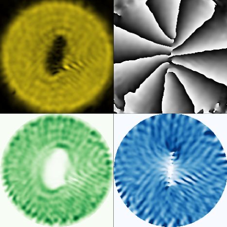
In this work we discuss how the classical orbital angular momentum (OAM) and topological charge (TC) of optical beams with arbitrary spatial phase profiles are related to the local winding density. An analysis for optical vortices (OV) with non-cylindrical symmetry is presented and it is experimentally shown for the first time that OAM and TC may have different values. The new approach also provides a systematic way to determine the uncertainties in measurements of TC and OAM of arbitrary OV.
arXiv , arXiv:1402.3226 (physics.optics) (2014).
15. Shaping optical beams with topological charge
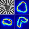
We show that by spatially arranging topological charges on a phase mask, it is possible to shape the spatial intensity profile of vortex beams in a controlled manner. As proof-of-principle experiments, we generated vortex beams with the spatial shape of straight lines, corners, and triangles. Potential applications for shaped beams include selective excitation of plasmonic modes, geometrically tunable Bose-Einstein condensates, and optical tweezers.
Opt. Lett. 38, 1579--1581 (2013).
